
��һ��
�Pע�ЈD�W
�ٷ���


������Ǖ�����>
-
>
�a�z�����Ʊ��[ (һ��2��)

-
>
�����W
-
>
(��)���ϹŴ��t�ҽ��
-
>
���t�䱾�Ď�Ӱӡ�cУ(��ذ�):�t��ժ�桤ѩ�����t���ϼ�
-
>
���t�䱾�Ď�Ӱӡ�cУ(��ذ�):��Ʒ����淽
-
>
���t�䱾�Ď�Ӱӡ�cУ(��ذ�):��ˎ���ɕ�
-
>
���t�䱾�Ď�Ӱӡ�cУ(��ذ�):����Ů��Ҫ�{��
�ЈD�r:¥137.2
����ُ��܇

�נ�����Ӱ��Ͳ���D�V �����Ϣ
- ISBN��9787547867822
- �l�δa��9787547867822 ; 978-7-5478-6782-2
- �b����105g���ۼ�
- �Ԕ������o
- ���������o
- ���ٷ��>
�נ�����Ӱ��Ͳ���D�V ������ɫ
�����·f���茍�������^���R�������rֵ�����^�i��ƺ����P���I�R���t����Ӱ��W���������t���ṩָ���͎�����
�נ�����Ӱ��Ͳ���D�V ���ݺ���
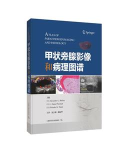
�����ɇ��H֪������ Alexander L. Shifrin ����F��������� 6 �����֣�չʾ�˸��NӰ��W�����ڼנ����ټ����\���еđ��ã�������Ҏ X �����߷ֱ��ʳ���CT ��MRI���Լ����t�W���g��ÿһ���D����Ԕ����עጣ�����������Y���c��׃�ą^�e�������x���R�e���ܵĮ��������x���s�IJ���׃�������^�i��Ƽ����P���I�R���t�����о����ṩ�˼נ����ٽ���֪�R�����������\������g�Ļ��A֪�R�c���M������������·f���茍�������^�ߵ��R�������rֵ�����^�i��ƺ����P���I�R���t����Ӱ��W���������t���ṩ��ָ���͎�����
�נ�����Ӱ��Ͳ���D�V Ŀ�
�� 1���� �נ����ٳ���sestamibi���輰����W
Ultrasound, Sestamibi Scan, and Pathology of the
Parathyroid Glands
��1�� �נ����ٳ� ������������������������������������������ 003
Parathyroid Ultrasound
Alexander L. Shifrin and Pritinder K. Thind
��2�� �נ����لӑB�@������������������������������������ 010
Scintigraphic Parathyroid Imaging
Pritinder K. Thind
��3�� �נ����ٲ���W ���������������������������������������� 015
Pathology of the Parathyroid Glands
Min Zheng and Virginia A. LiVolsi
��2���� �נ�������������������λ�ã�
����sestamibi����ʹ��w����W
Normal Anatomical Location of the Parathyroid Gland
Adenomas: Ultrasound, Sestamibi Scan, and Gross
Pathology
��4�� ���ϼנ����������������������������������������������� 035
Right Superior Parathyroid Adenoma
Alexander L. Shifrin and Pritinder K. Thind
��5�� ���¼נ����������������������������������������������� 043
Right Inferior Parathyroid Adenoma
Alexander L. Shifrin and Pritinder K. Thind
��6�� ���ϼנ����������������������������������������������� 056
Left Superior Parathyroid Adenoma
Alexander L. Shifrin and Pritinder K. Thind
��7�� ���¼נ����������������������������������������������� 066
Left Inferior Parathyroid Adenoma
Alexander L. Shifrin and Pritinder K. Thind
��3���� �נ��كȼנ��������������Լנ����������ͼנ����ٰ�
Intrathyroidal Parathyroid Adenoma, Cystic Parathyroid
Adenoma, and Parathyroid Carcinoma
��8�� ���z�顢sestamibi����� SPECT-CT sestamibi����
�נ��������������Լנ��������� ���������������������� 083
Ultrasonography, Sestamibi Scan, and SPECT/CT Sestamibi
Scan of Intrathyroidal Parathyroid Adenoma and Cystic
Parathyroid Adenoma
Alexander L. Shifrin and Pritinder K. Thind
��9�� �נ����ٰ���Ӱ��W�z�� ������������������������������ 107
Imaging of the Parathyroid Carcinoma
Alexander L. Shifrin, Pritinder K. Thind, Hubert H. Chuang, and Nancy D. Perrier
��4���� �נ������i��CT����
CT Scan of the Neck in Evaluation of Parathyroid Glands
�� 10�� �_չ�����о� ������������������������������������������ 119
Motivation for Imaging Studies
L. Daniel Neistadt
�� 11�� ����CT�ķ��� ���������������������������������������� 122
Contrast CT Approach
L. Daniel Neistadt
�� 12�� CT���g ���������������������������������������������� 125
The CT Technique
L. Daniel Neistadt
�� 13�� ���Ի���CT���� �������������������������������������� 127
Individual CT Phases
L. Daniel Neistadt
�� 14�� ����Ժͼ�����[��נ����ٵā�Դ �������������������� 130
Sources of False Positive and False Negative Enlarged
Parathyroid Glands
L.Daniel Neistadt
�� 15�� ���P�ij��z�� ���������������������������������������� 133
Correlative Ultrasound
L. Daniel Neistadt
�� 16�� �נ����ٵ��Π�����ʹ�С���������������������������� 135
Shape, Number, and Size of Parathyroids
L. Daniel Neistadt
�� 17�� �נ����ٵ�λ�� ���������������������������������������� 137
Location of Parathyroid Glands
L. Daniel Neistadt
�� 18�� ���ټ��� ���������������������������������������������� 142
Multigland Disease
L. Daniel Neistadt
�� 19�� ��Ҋ�ļנ����ٲ��Y������������������������������������ 144
Rare Parathyroid Presentations
L. Daniel Neistadt
��20�� ����չʾ ���������������������������������������������� 147
Illustrative Cases
L. Daniel Neistadt
��21�� �i��CT���茦�נ����ٵ��u�r ������������������������ 224
Summary: CT Scan of the Neck in the Evaluation of Parathyroid
Glands
L. Daniel Neistadt
��5���� �נ����ٶ�λ�Ľ����Լ��g
Invasive Techniques for Parathyroid Localization
��22�� �Є��Լ��g�נ����ٶ�λ ������������������������������ 229
Invasive Techniques for Parathyroid Localization
Richard Chang
��6���� �נ����ٵ�MRI�z��
MRI of the Parathyroid Glands
��23�� �נ����ټ�����MRI���� ������������������������������ 245
MRI for Imaging Parathyroid Disease
Jennifer L. Becker, Puneet S. Pawha, and Kambiz Nael
չ�_ȫ��
�נ�����Ӱ��Ͳ���D�V ���ߺ���
Alexander L. Shifrin�� �t�W��ʿ�����������ݺ����dzǝ���������W�t���������L. Daniel Neistadt�� �t�W��ʿ�������~�s�R�Z��˹ɽ����W��Pritinder K. Thind�� �t�W��ʿ�����������ݺ����dzǝ���������W�t�����ķ���ƌW ��־ɭ�������t����������λ���ڣ�����Ժ������N���ң���ʮ�����t�������Ѓ���W�Ǝ��^�ˣ��п�Ժ�������������T��������T�����F�Ό����ж����ʺ������\���������Ρ���������ʺ��Ʋ�ʿ�c���Tʿ�Wλ�cؓ؟�ˡ���������ʺ��ƌW���������Σ��Ї������t�Y�϶����ʺ��ƌW����ί���Ї������f���[�����ΌW����ί��ʡ�t�W�������ʺ��ƌ��I�W������ί��ʡ�����t�Y�όW�����Ǻ��Ʒ֕�����ί�����t�W�������ʺ��ƌ��I�W����ί�����¶����ʺ��^�i��ƌ��I30���꣬������g���s1000�_���ң������_չ�p���˹���ͬ�rֲ���ί����@������ؽ����ʡ������������P���B����ơ� ���ؽ���˯�ߺ�����ͣ���g�ί��ȡ��Ⱥ�����ʡ���n�}12헣��«@2025�ش�Ƽ����1헡��@ʡ�пƼ���17헣��Ե�һ����ͨ�����߰l��SCIՓ��36ƪ��Ӱ����Ӻ�Ӌ�s86.665���ڙ����Ұl������6헡����g�����������ж��ס����ж��R�����ʡ��ȡ����B��ʿ3�����Tʿ28����
�������]
- >
����Ԣ��-�����ČW�������-ȫ�g��

- >
���{����,��Ҫȥ��(2021�°�)
- >
�����c����ʿ
- >
�ƴ��Mʿ�
- >
�Ї��vʷ��˲�g
- >
�ҏ�δ��˾�����g
- >
������
- >
�Ї����ڞ��K��߅���^:�vʷ�c��W����
����N